Beyond the Routine: An Unexpected Obstruction in a Young Patient
Symptoms
An eighteen-month-old female Boerboel presented with an acutely bloated abdomen, persistent retching, and a severely diminished appetite just one week following a routine spaying procedure.
While initial blood work remained entirely within normal clinical limits, the sudden onset of these progressive gastrointestinal signs strongly pointed toward an acute structural blockage or significant fluid accumulation.
Visualisation
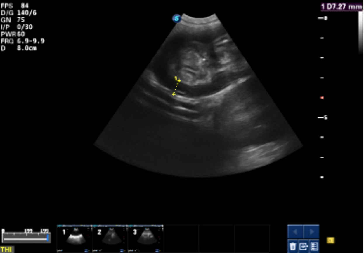
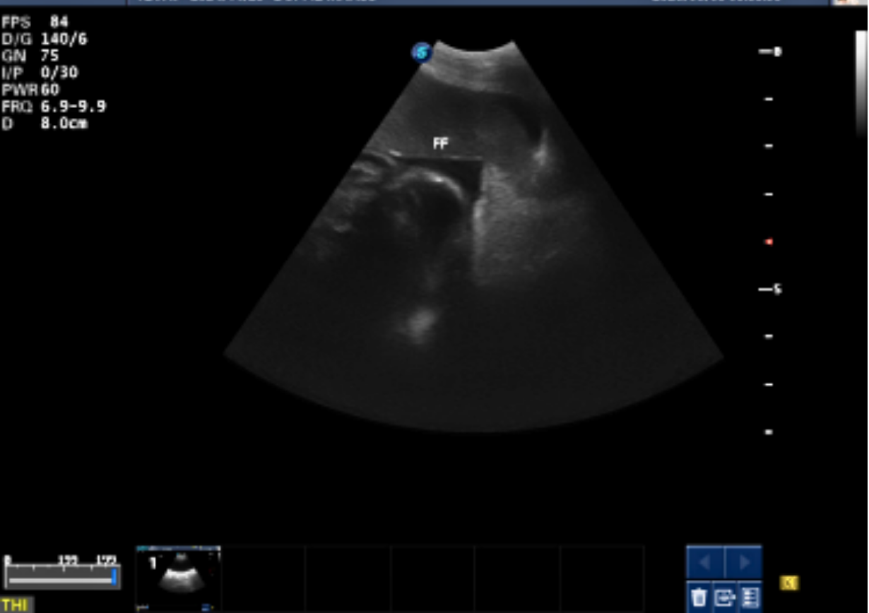
An extensive abdominal ultrasound was performed to thoroughly evaluate the gastrointestinal tract and surrounding organs, carefully mapping out the regional anatomy across multiple specialized views. To best illustrate this case visually, the optimal images to feature from the report are Figure 1 and Figure 2, which showcase the localized pyloric pathology clearly, alongside Figure 3 to demonstrate the peritoneal effusion
Figure 1
Figure 2
Figure 3
What it Revealed
The scan identified a severely thickened pylorus measuring 0.7 to 0.8 cm with a complete, highly concerning loss of its normal, distinct wall layering, alongside static gastric contents.
Secondary changes included mild free abdominal fluid, localised pancreatitis with surrounding mesenteric inflammation, regional lymphadenopathy, and a hyperechoic gallbladder wall, suggesting secondary cholecystitis.
Next Steps:
Immediate surgical exploration and full-thickness pyloric biopsies are essential to differentiate between severe mucosal hypertrophy and an infiltrative neoplastic process like lymphoma or adenocarcinoma. Supportive care consisting of gastroprotectants, antiemetics, and targeted nutritional adjustments should be maintained, with 3-view thoracic radiographs planned if histopathology confirms malignancy.
Important Lessons
Even in very young patients where benign chronic hypertrophic pyloric gastropathy is a primary consideration, a complete loss of gastrointestinal wall layering must always be treated with high diagnostic suspicion.
Mobile ultrasound acts as a vital tool for identifying silent secondary complications, such as localised pancreatitis and effusion, that fundamentally alter the immediate patient management plan.
About
"I dedicate my expertise to giving every pet the care they deserve."